
Cell atlas maps embryonic origins of human facial diversity
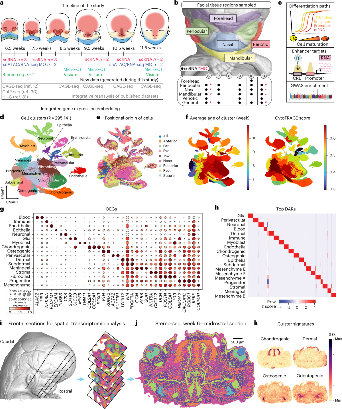
Researchers profiled single cells from human embryos between weeks 6 and 11 and identified 56 cell states tied to craniofacial variation, according to the paper in Nature Genetics. Gene expression links to facial traits were strongest in early mesenchymal progenitor cells, the authors found, before those links became restricted to specific regions. Peripheral nerves also fine-tuned the shape of the embryonic maxilla. Regulatory enhancers near pathology-related genes shaped facial diversity in modern human populations, and one enhancer tied to PAX1 expression proved important for normal skeletal development in mice.
Clinicians studying congenital craniofacial anomalies now have a reference map of the embryonic cell types affected by relevant genetic variants in humans and mice.
Written by the Genomes desk from the primary source cited and linked above and checked against it. Research use only; not medical advice. Corrections: [email protected].